
Clavicle Non Union Case
A 64-year-old gentleman, was referred for assessment of a painful right clavicle nonunion.
A 64-year-old gentleman, was referred for assessment of a painful right clavicle nonunion.
Peter is a 77-year-old man who first presented eleven years ago following a fall and injury to his right shoulder.

Dr Duckworth has published a research paper on how limited-incision plating is a safe and highly effective alternative for treating midshaft clavicle fractures.

I first saw Matthew who was a 20yr old professional skier when he had fallen off a trampoline whilst training and presented with a 3 week old displaced fracture of his clavicle which was in 2 main parts.

On the 14th of October 2024 a 53yo male presented with an acute rupture of his right distal biceps tendon after doing a deadlift competing overseas.

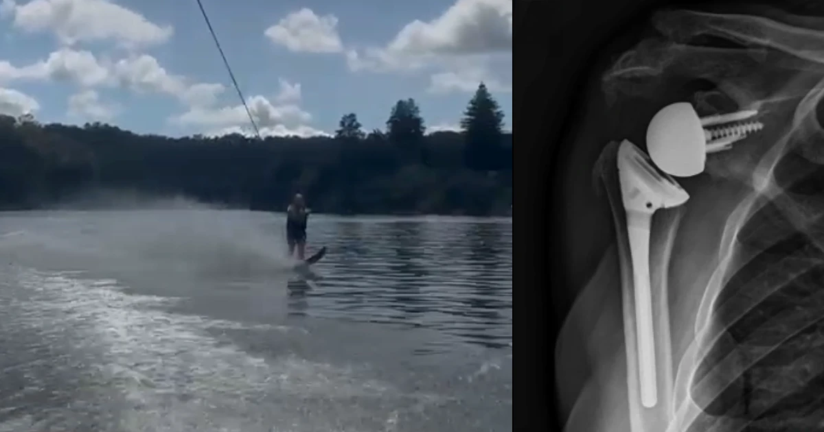
Tony is a 61 year old farmer, who lives in Rural NSW. His injury occurred when he fell off a horse and landed on his left shoulder.

Dr Duckworth has a published journal article investigating the functional outcomes of adolescent patients undergoing operative fixation of distal clavicle fractures.

This patient is a 67 year-old female who tripped and fell, landing on her right arm and breaking the top of the humerus.

This patient is a 14 year-old boy who injured his shoulder during a football tackle where his arm was forced into abduction.

Learn more about the stemless prosthesis used to replace the socket part of the shoulder joint during a primary shoulder replacement.

41 year old man who had broken his clavicle in a motor bike accident 20 years ago but has never felt right despite being 'fixed'.

This 76 year-old female suffered a significant fracture of her humerus after tripping and falling up a stair.

While lower limb fractures are more common in sport, upper body injuries including fractures of the clavicle or collar bone also occur.

Matt is a 26 year-old male who achieved improvement of his symptoms after surgery for a malunited clavicle fracture.

Joe is a 29 year-old male who had successful surgery to treat an old, malunited clavicle fracture that was causing pain and disrupting activity.

21-year-old professional mountain biker incurred a series of fractures to his collar bone, now well healed and back on the international mountain biking circuit.

This 38 year old man came off his motorbike at high speed on the race track and presented with a comminuted fracture of his clavicle.

A reverse total shoulder replacement is an orthopaedic procedure used to maintain the shoulder joint for people who have arthritis of the shoulder AND an irreparable, retracted rotator cuff tear.

Dr Duckworth summarises research data on the surgical treatment of midshaft clavicle fractures.

Collarbone fractures are common injuries in jockeys and track workers who are involved in riding and training horses.
For appointments and enquiries, please phone (02) 9806 3333
8am to 5pm, Monday to Friday
© 2008- Dr David Duckworth
Website by: ![]()